KindSIGHT Technology – The EIDON Retinal Camera & more
 Don’t be fooled by the fact that the KindSIGHT team members don’t sport nerdy black-rimmed specs – we love to geek out on the technology we have to offer! There are so many wild and wonderful instruments available when it comes to examining the eyes. We have carefully put together our toolbag of technology to create a suite of toys that are not only fun for us to play with, but intriguing for you to look at, and at the cutting edge of eye-examining inventions. Take a little tour through our collection of equipment to prepare yourself for your journey at KindSIGHT!
Say cheese with the EIDON Retinal Camera
Do you love a happy snap or 10? Then the EIDON is for you! This is a special camera that takes photos of the back of your eye, capturing your optic nerve, macula and a large area of your retina. It helps detect and monitor many conditions, including macular degeneration, glaucoma, freckles at the back of the eye, holes in the retina and much more! With the EIDON, it doesn’t matter what your best angle is, because it will capture all of them with its wide field of view. This machine has the capability to take multiple photos in each eye and stitch these together to form a single image, allowing us to see right out to the corners of your retina and detect any nasties that could be lurking in the shadows.
Another advantage of the EIDON Retinal camera is the ‘true colour’ imaging that it provides. We all know the wide variety of deceptive Insta filters available these days that leave pics looking nothing like the real thing. Some retinal cameras are similar, resulting in a big discrepancy between what the image captures, and what is really present. When it comes to looking at the back of the eye, we want the eye to show its true colours, and this is another area where the EIDON comes up trumps.
But wait – there’s more! The EIDON can capture pictures through small pupils, so you don’t even need the dreaded dilating drops in your eyes to have these images taken!
- EIDON retinal camera photograph
Looking Deeper – Heidelberg Spectralis OCT
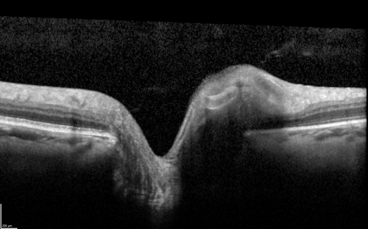
This little beauty is known as the ‘Rolls Royce’ of ocular coherence tomography (OCT) – say no more! In fact, NASA currently has the exact same model on the space station in order to understand why astronaut’s vision changes in space. Where the EIDON produces photos of the surface of the macula, optic nerve and retina, the OCT takes the investigation a step deeper. This technology shows what is happening beneath the surface of these structures, capturing a series of cross-sectional images and measuring the thickness of various tissues.
A key player in the detection and monitoring of many different conditions, the OCT is widely used to investigate glaucoma, macular degeneration, freckles at the back of the eye amongst a plethora of other pathology.
So what makes the Spectralis so speccy? Well, this instrument is particularly sensitive. It has the ability to take very fine measurements very quickly, meaning that if you move your eye, the machine can still record an accurate reading. The Spectralis also takes landmarks at the back of the eye and lines your eye up with these each time you have a scan performed to ensure that the exact same points are being measured across visits. Hence, we can be sure that we are comparing apples with apples, giving a high level of confidence when assessing for changes in the thickness of structures. This means better monitoring and management of conditions such as glaucoma and macular degeneration.
The Spectralis is so sensitive and specific to the patient, that it could be used for identification purposes. When performing repeat images, the machine will not function if the eye being tested does not belong to the original person who was scanned!
- A Spectralis OCT image of the macula
- A Spectralis OCT image of the optic nerve
The Side Vision Sidekick – Heidelberg Edge Perimeter (HEP)
The HEP and Spectralis OCT are like two peas in a pod. The HEP is a visual field’s machine, which measures peripheral vision. If you have never had the pleasure of performing a visual fields test, just ask someone who has how riveting this exam is. It involves pressing a buzzer every time you see a light flashing…similar to the most boring video game you have every played! However, this is a very important test largely used to examine for glaucoma. We can expect that any results we see indicating glaucoma on the OCT will correlate to those of the HEP, which makes these two a great little team!
Triple the fun – iTrace
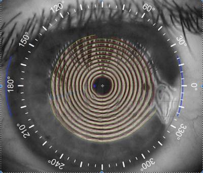
A three-in-one whiz-bang device, the iTrace is awesome at assessing a variety of conditions, including cataracts, pterygiums and any nasties that affect the shape of the front of the eye. It is a wavefront aberrometer (a fancy way of saying it measures the quality of vision), corneal topographer (creates a map of the front surface of the eye), and autorefractor (provides an estimate of your prescription in your glasses).
One of the most interesting features of the iTrace that patients enjoy is the function that demonstrates how a cataract is impacting on your vision. This device gives us a Dysfunctional Lens Index, which is a number on a scale of 0 to 10, with 10 indicating that the cataract is minimally interfering with your vision, and 0 indicating that the cataract is likely to be causing significant symptoms, such as blurred vision and glare. It also gives a lovely visual representation of how fuzzy the cataract is making your vision using a letter E. Sound interesting? Ask your doctor what your Dysfunctional Lens Index is!
The corneal topography feature takes a map of the front of the eye, not dissimilar to a topography map of mountains, where cooler colours show flatter areas, and warmer colours show steeper areas. Analysing the patterns helps to diagnose a variety of conditions that can affect the shape of the front of the eye, such as keratoconus, where the cornea is cone-shaped, or pellucid marginal degeneration, where the bottom half of the cornea is steeper. Identifying such abnormalities is important prior to proceeding with cataract or pterygium surgery to ensure the best outcomes are achieved.
- Dysfunctional lens index result in a patient without cataract
- Dysfunction lens index result in a patient with cataract
A Surgery Essential – the Lenstar
The Lenstar s also a star player in ensuring that our cataract patients have excellent vision following cataract surgery. The Lenstar is what we call a ‘biometer’, which takes measurements of the distances between the structures inside the eye, and the curvature of the front surface of the eye. These readings are used to calculate the best intraocular lens to use during cataract surgery. The accuracy of the pre-operative measurements that the Lenstar provides is extremely important in giving our patients the best shot at reducing their dependence on glasses after cataract surgery. Another nice feature offered by the Lenstar is the measurement of the thickness of the cornea, which gives us a more accurate measurement of intraocular pressure – very important in patients with glaucoma.
For those who have corneas thicker than average, the real pressure of the eye is lower than the amount measured. Similarly, the actual pressure is higher than the amount measured in those who have thinner corneas than average. If you have glaucoma or are being investigated for glaucoma, ask us what your corneal thicknesses are so you know if your readings need to be adjusted!
Who cares? iCare!
And what are we using to measure eye pressure? The i-Care! It’s no secret that the ‘puff of air test’ is everyone’s worst nightmare when having their eyes tested. However, having your eye pressure tested doesn’t have to leave you breaking out in a cold sweat, which is why at KindSIGHT we aim to be kind, with the iCare. This instrument measures the same thing as the ‘puff-of-air test’ but is far more comfortable to have performed – we think you’ll agree!
Last but not least – Lasers
There are a variety of purposes for lasers when it comes to the eyes. Our lasers at KindSIGHT are used in combination with a variety of high-tech lenses and special microscopes called slit lamps, to perform many painless procedures. These range from sealing holes in the retina to removing floaters and polishing the surface of lenses inserted during cataract surgery to reduce intraocular pressures in glaucoma.
We are very fortunate to have technology coming out of our ears – and eyes – at KindSIGHT. You can be confident that with our high level of training and state-of-the-art instruments we will provide you with a comprehensive and informative experience. If you would like to experience the KindSIGHT tech-tour first-hand, feel free to schedule a consultation with our friendly team.


