Ocular Coherence Tomography (OCT)
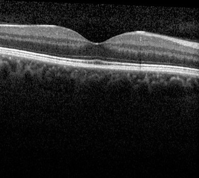
OCT is a technology that has changed the world of eyes dramatically over the past decade. While OCT is not a common household term, most people are at least familiar with an ultrasound. These two tests are very similar; the key difference being that OCT uses light waves instead of sound waves to create cross-sectional and three-dimensional images of various parts of the eye.
OCT scan
The OCT scan has allowed us to gaze deeper than ever before into our peepers, quite literally! Previously, we could only see with certainty what was happening on the surface of the retina and optic nerve. Thanks to the OCT scan, we can now also see what nasties may be lurking beneath the retina.
OCT scans also allow us to measure the thickness of various structures of the eye and monitor for very tiny changes over time. For eye health practitioners, this has improved our ability to track and monitor the progress of eye conditions more closely than ever before. Great news for our patients!
- ON THE SURFACE: A retinal photo showing the surface of the retina
- DIGGING DEEPER: An OCT scan showing a cross-sectional view of the retina
The Retina: A Layered Approach
If we think of the eye as a camera, the retina would be the camera film. Made up of receptors, the retina detects light signals coming from the images we see and creates a print of those pictures. Arranged across many different layers, the retina can now be seen in more detail than ever before, using OCT scanning.
There are many different conditions that can cause damage to specific layers of the retina. By being able to take an OCT scan, we can identify which layers are affected and more easily diagnose the condition. For example, many retinal pathologies can cause swelling, or a build-up of fluid, at the back of the eye. By monitoring the thickness of the retina and changes in the fluid bubbles, we can track if the condition is getting better or worse over time. This means we can assess if it is time to commence treatment, or if a current type of management is working.
- Cystoid macula oedema, showing fluid pockets at the macula
- The same macula following successful treatment!
Glaucoma: A bundle of Nerves
If we think of the eye as a camera again, the optic nerve is like the data cord: it transfers the pictures created by the receptors in the retina to your brain. Glaucoma is a condition that affects the optic nerve of the eye. In glaucoma, the tissue of the optic nerve gradually becomes damaged over time. It is often caused by high eye pressure and results in a loss of peripheral vision.
As glaucoma progresses, the nerve tissue becomes thinner and thinner, causing increasing vision loss. The good news is, treatment of glaucoma should significantly slow the progressive damage to nerve tissue.
To diagnose glaucoma, it is very important to monitor for changes in the nerve tissue over time. This is equally important to ensure the current treatment for a glaucoma patient is adequate. As you can imagine, it’s crucial that this analysis compares apples with apples. If we are not comparing the same point of nerve tissue across each visit, we cannot confidently say whether there has been a change. That’s where the ‘Rolls Royce’ makes its grand entrance.
The Heidelberg Spectralis OCT
At KindSIGHT, we are very lucky to have the Heidelberg Spectralis OCT, aka ‘The Rolls Royce’ of OCT imaging. This beautiful piece of technology takes images with very fine detail. This allows us to detect extremely fine changes over time and optimise the management of a range of eye conditions.
Here’s a fun fact – it is so technologically advanced that it is used by NASA for experiments conducted at their International Space Station!
The Heidelberg Spectralis OCT has the ability to take landmarks at the back of your eye and use these to make sure it is measuring the exact same points at each visit. Pretty impressive, right?
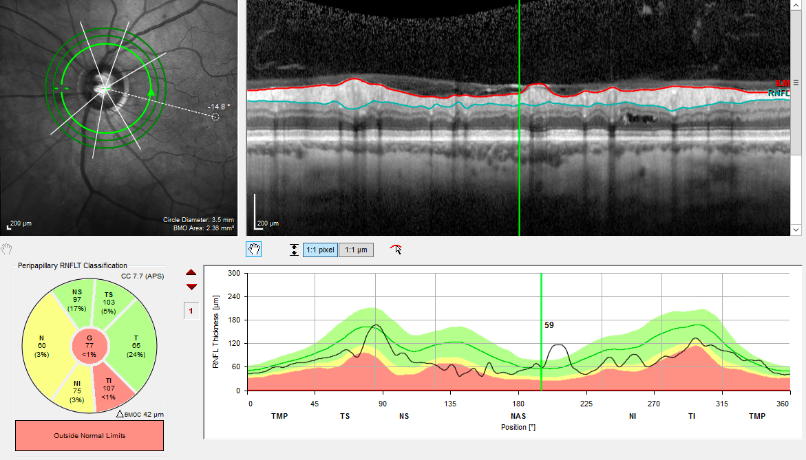
The Spectralis also has a large database of inbuilt information. This means it can compare the thickness of your nerve tissue against the rest of the population in your age group. This information gives us an accurate indication of whether your nerve tissue is thicker or thinner than it should be.
- An OCT scan of an optic nerve in a patient with glaucoma
- Three years later, OCT scanning in the same patient shows progressive thinning of the nerve, as the patient did not use the glaucoma drops prescribed to treat their glaucoma
But wait, there’s more!
So far we’ve explained how OCT scans examine the structures at the back of the eye. However, before we wrap up, it’s worth knowing one more thing. The Heidelberg Spectralis is in fact an all-rounder, in that it can also be used to scan the front of the eye!
In closed-angle glaucoma, for example, the angle at the front of the eye (the anterior chamber angle) is narrow, which prevents fluid drainage. This results in a build-up in eye pressure, which can then lead to glaucoma. The Spectralis OCT is able to take images of the angle at the front of the eye and diagnose whether it is narrow enough to lead to closed-angle glaucoma.
- An OCT scan showing a normal anterior chamber angle
- An OCT scan of the anterior chamber angle in a patient with closed-angle glaucoma
OCT scanning is a technology that the team at KindSIGHT is incredibly grateful for. As you now know, it helps us to diagnose and monitor eye conditions successfully for our patients over the long term.
If you have a condition you feel should be monitored by OCT scanning, or would like a baseline examination, contact us to schedule an appointment today!


